The common marmoset

For more information on the ecology, evolution, behavior and sociality of the common marmoset, visit Primate Info Net
scientific name:  Callithrix jacchus
common names:  Common marmoset, White-tufted-ear marmoset
distribution:  Brazil
height:  15-25cm (excluding tail)
weight:  250-550g 
life span:  12-15 years (average)
diet:  Exudative-insectivore
lifestyle:  Diurnal, arboreal
social organization:  Mixed-gender family units
conservation status:  Least concern

advantages of marmosets as model organisms for neuroscience research

The common marmoset has seen a rapid emergence as an ideal model organism for neuroscience and biomedical science over the last few years due to several physiological and logistical advantages. Their small size and ease of handling offers the convenience of laboratory rodent models such as rats and mice. However, unlike rodents, marmosets exhibit the shared physiological, behavioral and cognitive characteristics that are unique to primates, including the core functional architecture and organization of our nervous system. This unique complement of characteristics affords the exciting opportunity to feasibly utilize a primate species to model many of the diseases that afflict humans, ranging from those that affect humans at specific times in life – including both developmentally and during aging – to neuropsychiatric disorders that impact uniquely primate properties of our brain. Major advantages of marmosets for neuroscience research are outlined below.

small size and easy handling

The first advantage of marmosets over other NHP models such as macaques, is their small size, which is similar to lab rats. Adult marmosets are between 15 and 25cm high, excluding the tail, and weigh between 250 and 550 grams (obese animals may weigh more). These features make marmosets easier to handle than larger NHPs, requiring less cost and skill in their maintenance (eg. caging, feeding). Furthermore, due to their small size, marmosets require limited amounts of test compounds during experiments, a marked advantage when testing precious materials.

Easier handling is also attributed to the fact that to date, no fatal zoonotic diseases that can be transmitted to humans have been identified in marmosets, an advantage over macaques who can harbor herpes B.

High reproductive efficiency

Another advantage of marmosets is their high reproductive efficiency. Marmosets are easily bred in captivity and demonstrate the highest reproductive efficiency of any anthropoid primate. 

They reach sexual maturity at 1.5 years old and produce 2-3 offspring every 5-6 months. If an adult female is in breeding for 6 years, she will give birth about 12 times and will produce roughly 24 infants. 

Reproductive trait Rat Marmoset Macaque
Sexual maturity 10-12 weeks 1.5 years 3 years
Litter size 5-10 offspring 2-3 offspring 1 offspring
Gestation period 21-23 days 145 days 165 days
Delivery interval 2 months 5 months 1-2 years

As an example, 10 macaque breeding females will, in two years’ time, produce a maximum of 20 offspring, all of which will be immature (i.e. the reproductive population will not have increased during that time). In contrast, in the same 2-year period, 10 marmoset breeding females will produce an average of 60 offspring and the reproductive population will have tripled, as marmosets reach reproductive maturity by 14-18 months of age. 

Fast maturation and short life span

Marmosets offer unique opportunities to study and understand biomedical processes that have not been feasible to model in other NHPs. The fast maturation and relatively short life span of marmosets makes them a valuable resource to study developmental, chronic and aging related disease, which are among the most pressing US human health concerns. Marmosets reach sexual maturity at around 1.5 years and by 8-13 years of age show signs of age-related pathologies such as an increase in cancers, amyloidosis, pathogenic tau accumulation, diabetes and renal diseases, a decrease in lean muscle mass, hearing loss as well as declines in cognitive and motor function with increased age. 

Notably, the time period required to move from early to late life (i.e. to advance through the stages of development, aging or chronic disease) is within the range of a typical, NIH-funded project. Macaques on the other hand do not reach sexual maturity and geriatric age until around 3 and 20 years, respectively. 

Furthermore, like humans, marmosets are typically group-housed with pair-bonded parents and multiple litters of offspring, making it possible to observe the process of aging across two generations of animals in the same cage. This is a particularly attractive feature for transgenic models in which disease onset may depend on aging factors, such as neurodegenerative disorders like Alzheimer's Disease and Parkinson's Disease. 

brain anatomy and function

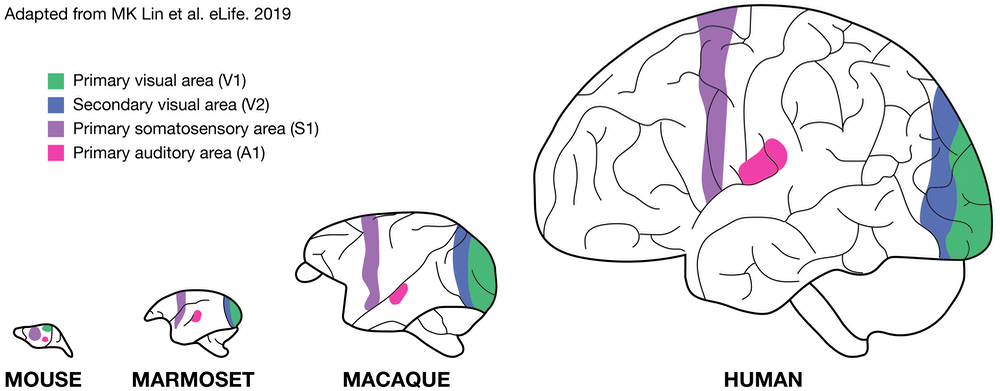
Although the marmoset brain is small, it retains anatomical and functional organization specific to primates, including humans. This includes a large neocortex, granular prefrontal cortex, expanded visual and auditory cortical fields and reduced olfactory regions. Like humans, the brain of marmosets have a large amount of white matter, a feature that makes them promising as a translational model of brain disease. The marmoset brain is also well suited for studying circuit wiring and connectomics in a complex NHP because it does not pose the same big data challenge for computational tools as the macaque brain which is several magnitudes larger. 

In addition, in contrast to the large and convoluted macaque brain, the marmoset cortex is lissencephalic (i.e. smooth) like a rodent's. This greatly facilitates the mapping of functional brain areas by neuroimaging techniques, such as fMRI and optical imaging, as well as by electrophysiology, with high spatial resolution as all of the neocortical fields are accessible directly below the skull rather than within sulci. Although it is important to note that the lissencephalic brain is dissimilar from humans. 

comparable cognitive characteristics to humans

Primates are distinguished from other animals by the breadth and sophistication of our sociality, including the dynamic models we develop for social decision-making to effectively navigate the complexities of these social landscapes. While marmosets share these attributes with other primates, their societies also exhibit characteristics that are typical of humans yet rare in other primates.

Marmosets, for example, pair-bond and cooperatively care for their offspring. Cooperation occurs frequently in marmoset society and the species displays high levels of prosociality. Several experiments show that marmosets also utilize imitation, a distinct social learning mechanism that is rare amongst primates as it had previously only been reported in humans and chimpanzees. 

Marmosets are also highly vocal, engaging in near tonic levels of conversational exchanges with conspecifics by utilizing turn-taking mechanisms that are learned during development. While vocal communication is critical to mediating their social interactions, marmosets also utilize diverse visual signals – including facial expressions – as a parallel social signaling system, similarly to humans.

Furthermore, marmosets have the ability to perform cognitive tasks similar to those often used in human studies, such as visual discrimination and reversal learning.

RESEARCH techniques in marmosets

NEUROimaging

Neuroimaging techniques are available to study anatomical structures and functional processes in the brain as well as to better understand the mechanisms underlying brain pathologies. The lissencephalic brain of the marmoset is a significant advantage, facilitating a wide-range of neuroimaging studies. The small brain size makes large-scale manipulation of cortical activity feasible, which is useful for studying inter-area interactions. 

Anatomical mapping of the marmoset brain is a mature and active area, and multiple high resolution 3D MRI atlases are available (BMA, marmoset brain mapping). Connectivity atlases are being constructed using fluorescent retrograde tracer injections to map the connections between different cortical areas (marmosetbrain.org).

Functional MRI (fMRI) is a powerful technique for allowing areal observations of functional brain activity and it is the main research tool in cognitive neuroscience. In marmosets, fMRI has been used to study various sensory systems, including somatosensory, auditory and visual pathways. Resting-state studies have been used to study functional brain networks and show promise as a possible diagnostic tool for brain disorders in humans.

Although powerful tools, MRI and PET do not have the resolution to investigate neural events at the cellular level. Light-based imaging techniques such as confocal or two-photon microscopy are complementary methods to visualize processes in the brain and can offer the spatial and temporal resolution that is required to study individual cells or cell populations in the brain. Specifically,  genetically encoded calcium indicators (GECIs), such as GCaMP have been used to visualize subcellular, cellular and ensemble neural dynamics in the marmoset brain. These indicators can be delivered to discrete areas of the brain via stereotaxic injections of recombinant adeno-associated viruses (AAV) or lentiviral vectors. Alternative to virus-mediated transgene expression is the development of transgenic animals lines that endogenously express GECI molecules in the brain.

While GECIs provide a readout for neuronal activity, optogenetic tools allow for the very specific control of neuronal activity. Optogenetics is a rapidly growing field with a rapidly growing toolkit, but the basic premise is to use genetically engineered opsin proteins to selectively manipulate target cells using light. Optogenetic cortical stimulation has been demonstrated in marmosets (e.g. T Ebina et al. 2019). Finally, new AAV vectors for delivering GECIs and optogenetic constructs are actively being designed to have highly specific trophism to a wide variety of marmoset brain cell types (e.g. NC Flytzanis et al. 2020).

transcriptomics

A number of gene expression/transcriptomics projects are underway in marmosets, particularly with regard to the brain.
For example, the Marmoset Gene Atlas provides an in situ hybridization-based database of genome-wide, high-resolution  gene expression throughout the marmoset brain.

Furthermore, single cell RNA sequencing technology coupled with multiplexed error-robust in situ hybridization (MERFISH) is now being applied to marmoset brain transcriptomics to identity and spatially map cell types across multiple regions of the adult marmoset brain (BICCN U01 Feng). 

marmoset genomics

The first draft of the marmoset genome was published by the Marmoset Genome Sequencing and Analysis Consortium in 2014 and the female animal became the first New World monkey to have its complete genome sequenced. Currently, there are 12 assemblies available. The genome size is 2.9 Gb and contains 42,294 genes of which 52.8% are protein-coding. Ongoing sequencing projects continue in order to improve the existing genome assemblies with the goal of increasing the efficiency of genome research, transcriptome analysis and the generation of genetically modified marmosets.

Additionally, the MCC is committed to generating whole genome DNA sequence data for the entire population of research marmosets in order to effectively manage the genetic composition of the existing US marmoset population, avoid inbreeding and maximize the research utility of these animals. An important consideration for these sequencing studies is the source of the DNA. Marmosets predominantly produce dizygotic twins sharing a single placenta. This unique feature results in exchange of hematopoietic stem cells between litter mates and lifelong blood chimerism. This chimerism complicates the collection of germline DNA from blood for genome sequencing because a high fraction of circulating blood cells are derived from not the animal bled, but from their non-identical twin. The extent of chimerism varies widely across tissues from blood (~50%) to skin (~10%) and hair follicles (1%). 

genetic modification and genome editing

The high-fertility of marmosets is a specific advantage for technologies, such as transgenics, in which rapid establishment of genetically defined lines is essential. While application of transgenic technologies to NHPs will likely remain expensive when compared to rodents, the use of marmosets brings this technology within an acceptable financial range for applications in neurodegenerative diseases, for example, where NHPs are the most appropriate models. 

Genetically modified marmosets have been successfully generated by simple viral transgenic strategies as well as via genome editing with site-directed nucleases. The first successful demonstration of germline transmission of a transgene in NHPs was done in marmosets by Sasaki and colleagues in 2009 via injection of a lentiviral vector carrying green fluorescent protein. Since then, several transgenic marmosets have been generated using this technique and efforts have accelerated to improve the implementation of transgenic technologies in marmosets. New viral based approaches now make it possible to more selectively target specific cell types and circuits in the brain. These viral technologies include the intravenous delivery of adeno-associated virus (AAV) capsids designed to cross the blood-brain barrier in marmosets. 

Modern genome editing technologies using site-directed nucleases such as zinc finger nucleases (ZFNs), transcription activator-like effector nucleases (TALENs) and the CRISPR/Cas9 system (clustered regularly interspaced short palindromic repeat/CRISPR associated 9 are now being applied in marmosets, particularly to generate target gene knock-out/knock-in animals.

Another potential approach for making genetically modified animal is to perform gene modification in embryonic stem cells (ESC) or iPSC followed by selection of appropriately modified stem cells via genetic screening which will then develop into chimeric gene edited offspring. 

Disease models in marmosets

Marmosets are utilized as animal models in a wide diversity of biomedical research disciplines, ranging from infectious disease (eg. Zika, Hepatitis, E. bola) to reproductive biology but neuroscience remains the dominant area in which this New World primate is used. Some of these disease models include the following:

  • Aging
  • Alzheimer's Disease
  • Parkinson's Disease
  • Huntington's Disease
  • Multiple Sclerosis
  • Stroke
  • Spinal cord injury
  • Major depressive disorder
  • Anxiety
  • Behavioral endocrinology
  • Hearing loss
  • Vision
  • Obesity
  • Toxicology
  • Metabolic disease